Mom Had One

Years ago, when I was a young woman in my 20s, my mother had a fibroid in her uterus. Or, at least that was the way she explained it. Come to think of it, she didn’t explain. Rather, she simply announced she’d had it removed. My brother and I, both medically ignorant at the time, had no idea why that was such a big deal.

In the intervening decades, I’ve learned a lot. Most of that medical knowledge has come from researching for the blog. That’s how I learned that a fibroid is a tumor or, as Johns Hopkins puts it:

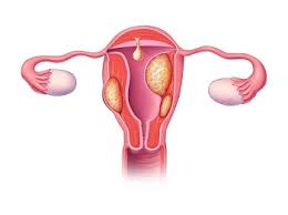
“Fibroids are growths made of smooth muscle cells and fibrous connective tissue. These growths develop in the uterus and appear alone or in groups. They range in size, from as small as a grain of rice to as big as a melon. In some cases, fibroids can grow into the uterine cavity or outward from the uterus on stalks.”

Notice the word tumor wasn’t used in this definition. It didn’t have to be because a tumor as defined by my all-time favorite dictionary, the Merriam-Webster, is:

“an abnormal benign or malignant new growth of tissue that possesses no physiological function and arises from uncontrolled usually rapid cellular proliferation”

Since they have no function and grow in the same way as cancer does, are they cancerous? Not according to Planned Parenthood, who also offers us the symptoms:

Photo by Sora Shimazaki on Pexels.com

“Uterine fibroids are almost never cancerous, and they don’t increase your risk for getting other types of cancer. But they can cause pelvic pain, heavy period bleeding, bleeding between periods, back pain, and in some cases, infertility or miscarriages. However, many people with fibroids don’t have any symptoms at all.”

My mother was not the type to want to know how the fibroid developed. As most people did 50 years ago, she just wanted it gone. But you might want to know. WebMD explained:

“Experts don’t know exactly why you get fibroids. Hormones and genetics might make you more likely to get them.

HormonesEstrogen and progesterone are the hormones that make the lining of your uterus thicken every month during your period. They also seem to affect fibroid growth. When hormone production slows down during menopause, fibroids usually shrink.

Genetics. Researchers have found genetic differences between fibroids and normal cells in the uterus.

Other growth factors. Substances in your body that help with tissue upkeep, such as insulin-like growth factor, may play a part in fibroid growth. 

Extracellular matrix (ECM). ECM makes your cells stick together. Fibroids have more ECM than normal cells, which makes them fibrous or ropey. ECM also stores growth factors (substances that spur cell growth) and causes cells to change.”

Let’s get back to Mom wanting it gone. The question here is how? It turns out there are many, many different methods from different types of ablations, surgeries, and medications.

I know you want to know what this has to do with chronic kidney disease. That is actually what I wanted to know, too. According to The National Library of Medicine:

“Uterine fibroids constitute the most common tumor in women of reproductive age …. Significant morbidity secondary to fibroids is a rare event; however acute complications from fibroids may include thromboembolic events, acute torsion of pedunculated fibroids, acute abdominal pain, vaginal bleeding, intra-abdominal bleeding, acute urinary retention, and renal failure. [Gail here: I bolded that.] Uterine fibroids are associated with obstructive renal failure as they can physically compress the ureters, leading to acute urinary retention and postrenal nephropathy.”

I get it, but it took me a while to figure out what this meant. So I looked for a different, more easily understood  explanation… and found it on Fibroids.com:

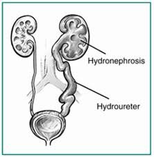
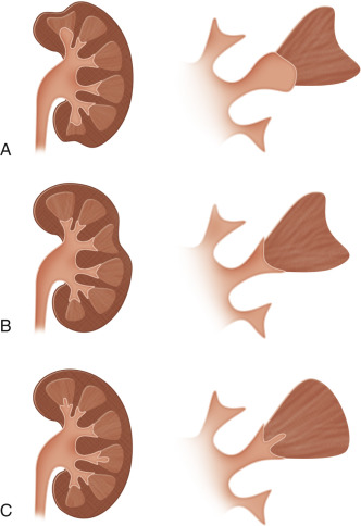
“Although fibroids are made of muscle tissue found in the uterus, they can outgrow the space within the uterine walls and expand to a size large enough to affect the ureter. The ureter is the tube that connects the bladder and the kidney. When fibroids down [sic] on the ureter, the kidneys swell and develop a condition known as hydronephrosis.

Hydronephrosis is often associated with painful urination, an increased urge to urinate, as well as flank and back pain. In more severe cases, permanent kidney damage may also occur. If you are currently experiencing any of these symptoms or suspect your kidneys may be at risk due to your fibroids, consult with your doctor immediately.” 

Talkingfibroids,com tells us more about hydronephrosis:

“But if hydronephrosis persists for a long time, the nephrons (kidney cells) can die, and the result can be irreversible kidney damage. Even if the obstruction to the ureter is eventually removed, a kidney that has gotten to this point will not regain function.”

Hmmm, there must be a way to prevent this. I searched and searched until I found what I was looking for on India’s GAURI – Guna Associates in Urogynecology & Research for Incontinence:

“The removal of fibroids is crucial for those suffering kidney complications due to fibroids. Despite the prevalence of fibroid surgery like a hysterectomy or myomectomy [removal of only the fibroid], there is a less invasive procedure called uterine fibroid embolization (UFE) that eliminates the scars and trauma associated with surgery.

Fibroid embolization works by reducing the larger fibroid that is pushing on the ureter and causing kidney problems. It provides a quick and effective procedure with no chance of regrowth of the fibroid. By shrinking the fibroid instead of removing it, patients experience a quick and effective procedure.”

While that may sound scary, remember that surgery is another way to deal with fibroids but UFE is less invasive. There is also medication, but please do not take NSAIDS. That stands for non-steroidal anti-inflammatory and, as CKD patients, is not for us. And let’s not forget ablation.

As for diagnosing hydronephrosis, the usual blood and urine tests plus an ultrasound does the trick. The ultrasound will let you see if the kidney is swollen. The urine test will rule out infection or urinary stones. And the blood test will evaluate your kidney function. I wonder whether Mom underwent these tests.

Until next week,

Keep living your life!

More Needling

Last week’s blog about biopsies didn’t feel complete to me. Comments from readers indicate they felt the same way. This week, I thought I’d explore some of the issues they asked about.

The question most often asked was, “Does it hurt?” This is a tricky one since each person is unique. Let’s see what Mount Sinai had to say,

“Numbing medicine is used, so the pain during the procedure is often slight. The numbing medicine may burn or sting when first injected.

After the procedure, the area may feel tender or sore for a few days.

You may see bright, red blood in the urine during the first 24 hours after the test. If the bleeding lasts longer, tell your provider.”

Some readers have reported that they had, indeed, experienced soreness – but not necessarily pain – for several days after the procedure. On a personal note, I can sometimes have soreness from my insulin injections. Again, people are different.

Another reader wanted us to know that it was possible for a kidney biopsy to go wrong. I turned to Johns Hopkins to see what I could learn,

“As with any procedure, complications can happen including:

  • Bruising and discomfort at the biopsy site
  • On-going bleeding from the biopsy site, in the urine, or inside the body
  • Puncture of nearby organs or structures
  • Infection near the biopsy site

If the kidney biopsy is done with the aid of X-ray, the amount of radiation used is small. Therefore, the risk for radiation exposure is low.

If you are pregnant or think you may be, tell your healthcare provider. Talk to your healthcare provider about the risks to the fetus from being exposed to an X-ray. Pregnancy is not always contraindication for having a kidney biopsy. It may be important to maintain the health of the mother. Special precautions may be taken to protect both the mother and the fetus during a kidney biopsy.

You may not be able to have kidney biopsy if you have an active kidney infection, certain bleeding conditions, uncontrolled high blood pressure, or have only one working kidney.

There may be other risks depending on your specific medical condition. Be sure to raise any concerns with your healthcare provider before the procedure.”

I believe the reader in question had an infection. I can’t stress enough that although these readings report what most people experience, each of us is unique, different, and may have slightly different reactions.

As far as preparation for a kidney biopsy, The National Kidney Foundation covered that one:

“For most kidney biopsies, patients are advised to not take over-the-counter pain medicines such as aspirin, ibuprofen (Advil®, Motrin®), naproxen (Aleve®), or other medicines that may cause thinning of the blood for 2 weeks prior to the test. These medicines can change the way the blood clots and raise the risk of bleeding. For the same reason, you will likely be instructed to stop taking certain supplements such as fish oil.

Blood and urine samples are usually taken before the kidney biopsy to make sure you do not have an infection or other condition. Your doctor may also want you to change other medications before the biopsy. You may be told you should not eat or drink for eight hours before the procedure.”

I thoroughly enjoyed their use of the word ‘most’. However, I did wonder why a kidney patient would be taking NSAIDS [Nonsteroidal anti-inflammatory drugs] in the first place since they can harm your kidneys, even your transplanted kidneys.

I wasn’t that clear about the connection between bleeding and fish oil. Never fear! Mayo Clinic to the rescue,

“While generally safe, getting too much fish oil can increase your risk of bleeding and might affect your immune response. It’s not clear whether fish oil is safe for people who are allergic to seafood. Take fish oil supplements under a doctor’s supervision.”

All right, what have I left out? Let me think for a moment. Of course! Some readers wanted to know if they needed to be hospitalized for a kidney biopsy. Interestingly, some will. But most people will just need to be observed for a few hours before they go home to rest for a day or two.

I kept reading that the actual procedure takes only 15 minutes but with the preparation and the immediate after care, it takes an hour. Something else that was repeated on site after site was that that it’s preferred you don’t drive for a day or two after the procedure or operation.

A procedure refers to a medical intervention that doesn’t break the skin, while an operation does. For example, a closed [needle] biopsy is a procedure while an open biopsy is a surgery. Also, most readers thought a kidney biopsy was only used to evaluate the health of a kidney transplant. The Cleveland Clinic explained that there are other uses for a kidney biopsy,

“A kidney biopsy helps doctors identify the cause of kidney problems so they can treat the condition effectively. It can reveal scarring, inflammation (swelling), and protein deposits that cannot be identified with other tests, such as ultrasounds or blood and urine tests.”

Unfortunately, it can take anywhere from two to ten days to receive the results of the biopsy. It depends upon how many tests were being performed on the sample. As for whether or not you can go to the toilet after the test, the answer is no. You’ll be handed a bed pan. That’s so the doctor and/or nurse can see if you’re passing blood in your urine and, if so, how much. You’ll probably receive a blood transfusion if your overall blood loss is deemed too much.

I think I’ve covered everything I didn’t in last week’s blog. I urge you to remember that each patient is unique. That’s what is meant by ‘precision medicine.’ Your doctor will be the best person to make these decisions and discuss them with you.

Until next week,

Keep living your life!

This Nutcracker is Not the One in the Nutcracker Suite

Today is my second grandson’s first birthday. So, I thought I’d look for some music he might like. There was always The Nutcracker Suite, I thought. Although it was written in 1892 by Tchaikovsky, it’s been a favorite of children everywhere ever since. I’m guessing that’s due to the ballet.

But I’m not writing about that nutcracker. I’m writing about nutcracker syndrome today. I promise this is not a joke. There is such a condition. This is the first time I’ve heard of it even though I’ve been writing about all things kidney since 2010, so don’t feel bad if you’ve never heard of it.

Let’s start at the beginning with a definition. One of my trusted sites, the Cleveland Clinic, explains it this way:

“Nutcracker syndrome is a condition that affects your left renal vein. This is the vein that carries blood away from your left kidney and back to your heart. Nutcracker syndrome is a type of extrinsic vein compression syndrome. In these syndromes, the structure of your blood vessels puts pressure on one of your veins.

If you have nutcracker syndrome, two arteries in your belly compress part of your left renal vein. This compression raises the blood pressure in your renal vein and forces some blood to flow in the wrong direction. As a result, nearby veins swell, causing symptoms and potentially leading to complications.”

Frankly, this sounds painful. I didn’t know which to explore first, symptoms or causes. I chose alphabetically. Remember, I was an English teacher for many, many years. So, causes it was.

India’s Icliniq, a second opinion site, offered both causes and risk factors:

“The specific causes of nutcracker syndrome vary from person to person. Sometimes, people are born with variations in the blood vessels that can result in the symptoms of nutcracker syndrome. The symptoms of nutcracker syndrome are commonly seen in females in their 20s and 30s. The factors that increase the risk of nutcracker syndrome are listed below:

  1. Pancreatic tumors increase the risk of nutcracker syndrome because the vessels supplying the pancreas become damaged and compress the renal veins, resulting in nutcracker syndrome.
  2. Curvature in the lower spine.
  3. Tumors in the tissues present in the inner lining of the abdominal wall.
  4. Nephroptosis (a condition in which the kidneys drop into the pelvis while standing).
  5. Aneurysm in the abdominal aorta.
  6. Sudden changes in the weight and height.
  7. Enlargement of abdominal lymph nodes.
  8. Pregnancy.”

I’d had a pancreatic tumor, but I didn’t develop nutcracker syndrome. I guess that makes me one of the lucky ones. I constantly marvel how we, as humans, find the good in everything: pancreatic cancer? Bad. No development of nutcracker syndrome: good. By the way, I also have curvature in the lower spine, but that doesn’t seem to be affecting anything.

Time to find out the symptoms now. MedicalNewsToday had that covered:

“The symptoms of NS [nutcracker syndrome] can vary depending on the extent of LRV [left renal vein] recompression. Some people do not experience any symptoms.

When symptoms occur, they may include:

Wait a minute. Flank pain, protein in the urine, and high blood pressure may also be signs of chronic kidney disease. Is nutcracker syndrome considered a kidney disease? After a little digging, I found it is, indeed, a kidney disease although a rare one.

Since it is a rare disease, I wondered how it was diagnosed. Healthline to the rescue:

“First, your doctor will perform a physical exam. Next, they’ll take a medical history and ask about your symptoms to help them narrow down a possible diagnosis.

If they suspect nutcracker syndrome, your doctor will take urine samples to look for blood, protein, and bacteria. Blood samples can be used to check blood cell counts and kidney function. This will help them narrow down your diagnosis even further.

Next, your doctor may recommend a Doppler ultrasound of your kidney area to see if you have abnormal blood flow through your veins and arteries.

Depending on your anatomy and symptoms, your doctor also may recommend a CT scan or MRI to look more closely at your kidney, blood vessels, and other organs to see exactly where and why the vein is compressed. They might also recommend a kidney biopsy to help rule out other conditions that can cause similar symptoms.”

Let’s say [heaven forbid] that you or someone you love is born with or develops nutcracker syndrome. What can be done about it? Thank you to UCLA Health for the following:

“Patients with mild symptoms may be observed. Children may be given time to grow and weight gain can help others. For patients with severe symptoms, one of several procedures may be recommended based on anatomy, symptoms, age, and odds of symptom relief…. 

  • Open or laparoscopic/robotic surgery involving repositioning of the renal vein in a way that frees it from compression (renal vein transposition)
  • Open or laparoscopic/robotic surgery involving bypass of the compressed renal vein
  • Autotransplantation of the left kidney to the pelvic vessels
  • Endovascular approaches including placing a stent in the left renal vein”

I can’t help but wonder what the outcomes could be. After all, this is a rare disease. I turned to Orphanet for an answer. This is the portal for rare diseases and orphan drugs.

“As this is a benign condition, overall prognosis is excellent. In highly symptomatic patients, with severe pain, frank/recurrent hematuria requiring blood transfusion, active intervention needs to be considered. Prognosis following intervention is excellent.”

For those of you wondering, this is not a disease for which a change of lifestyle will help.

Now comes the information you’ve been waiting for. Why is it called nutcracker syndrome? We all know what a nutcracker looks like, right? Imagine a nutcracker squeezing your left renal vein. Ouch!

Until next week,

Keep living your life!

Kidney Diffuse Parenchymal Disease Bilateral

Yep, that was my reaction too when a reader asked me what this was. I took a bunch of guesses and then asked her to please speak with her nephrologist about this. Sometimes, I get asked doozies. I turned to the National Kidney Foundation for a simple explanation of this condition:

“’Bilateral renal parenchymal disease’ is a doctor term for scarring changes in the substance of both kidneys.”  

I did look for a simple explanation, but that’s too simple for me. For instance, what are these ‘scarring changes’?

Oh, wait. Dr. Prashant C Dheerendray at Dharma Kidney Care in Bangalore, India, tells us something we should be aware of before we start investigating anything about this condition:

““Renal parenchymal disease” is a term used to describe the appearance of the kidneys on ultrasound. It doesn’t give the complete information about the functioning of kidneys in a given patient. Hence, as a nephrologist, I need more information from blood and urine tests before deciding whether it is dangerous or not.”

Time to hear from my favorite dictionary for some help:

of, relating to, or affecting the right and left sides of the body or the right and left members of paired organs”

That makes perfect sense since bi is from Latin and means two, while lateral is also from Latin and means side. Most people have one kidney on each side of their body. Hence, bi for two and lateral for sides. Many thanks to the Merriam-Webster Dictionary, as usual.

Uh-oh, we forgot ‘diffuse.’ Back to the dictionary:

“spread or cause to spread over a wide area or among a large number of people”

I suspect including diffuse means the scarring is spreading. We’ll find out in just a little bit. So far, I’ve defined two not necessarily medical words. We know that renal and kidney are the same, so we’re left with ‘parenchymal.’ It does remind me of the term for an elephant, but I kind of doubt that’s the case here. We’re really working the dictionary today:

“relating to or affecting the functional tissue of an organ”

Oh, that’s where the scarring comes in. Let’s see if we can figure out exactly how, though.

Healthmatch, according to their website, is:

“… a diverse team of doctors, engineers, scientists and people dedicated to challenging the status quo of medical research.

We are united by a passion to deliver better healthcare options, for all, regardless of location, background or means. This means access to trials and the revolutionary treatments that come from them.”

 It tells us:

“The kidneys comprise various components and structures that contribute to their bodily function. Within the kidney’s anatomy is the parenchyma, which is responsible primarily for the filtration of blood that passes through the kidneys and the excretion of waste in the form of urine.”

So, the scarring interferes with the blood filtration and excretion of waste, two of the most primary of the kidney’s many functions. Let’s see if can figure out how this scarring happens. I lucked on to Nicklaus Children’s Hospital’s information about the parenchyma:

“The renal parenchyma is the functional part of the kidney that includes the renal cortex (the outermost part of the kidney) and the renal medulla.

  • The renal cortex contains the approximately 1 million nephrons (these have glomeruli which are the primary filterer of blood passing through the kidney, and renal tubules which modify the fluid to produce the appropriate amount/content of urine).
  • The renal medulla consists primarily of tubules/ducts which are the beginning of the collecting system that allows the urine to flow onwards to being excreted.

Renal parenchyma disease describes medical conditions which damage these parts of the kidney.”

Now that I know what this condition or disease is, I’d like to know what causes it. I,Cliniq, the Virtual Hospital to the rescue!

“The causes of the renal parenchymal disease include:

We don’t know what the ‘others’ are, but I was surprised to see two non-kidney causes in this list: ‘bacterial and viral infections’ and ‘drug- related’. This is starting to sound like something you want to deal with a.s.a.p. But how?

According to Healthmatch:

“Doctors offer no single common solution or plan for the management and treatment of renal parenchymal disease. Each approach to helping an individual with renal parenchyma disease will take into account their condition, what symptoms they are experiencing, and the severity of the damage to the renal parenchyma.

The reality is that there is no cure for renal parenchyma disease but rather medical management of the symptoms as best as possible to try and prevent further deterioration and damage to the kidneys and your overall health.”

No cure? We’re living with chronic kidney disease which has no cure, but we can slow down its progress and do our best to keep it from being dangerous. Is renal parenchyma disease dangerous? Is CKD dangerous?

The National Kidney Foundation has a revelation for us:

“Renal parenchymal disease means the same thing as chronic kidney disease (CKD).  It is just another way of saying CKD.”

Everything you’ve learned about CKD is true about renal parenchymal disease bilateral, too. You can call me Gail, Mom, or Bubby, but I’m still the same person. You can call this disease renal parenchymal disease bilateral or CKD, but it’s still the same disease.

Until next week,

Keep living your life!  

These are not the Lucky Kind 

We all know about the superstition that horseshoes are lucky. Some might even argue that this is fact, not a superstition. Today, I’ll be writing about the unlucky kind of horseshoe. Some of you may know about this already; some may not. I didn’t and was surprised by what I found. 

First off, a definition of horseshoe kidneys [Surprise!] might be helpful. WebMD was pretty comprehensive in their definition: 

“Horseshoe kidney, also called renal fusion, is a condition that starts before a child is born. 

As a baby develops in the womb, their kidneys move into position just above the waist — one on each side of the body. But sometimes that doesn’t happen as it should. Instead, the kidneys fuse together at their base, forming a U or horseshoe shape. It usually happens between weeks 7 and 9 of the pregnancy. 

The condition isn’t common — about 1 in 500 babies have it, boys more often than girls. And many kids won’t have serious health issues because of it. 

However, about one in three children with fused kidneys will also have a problem with their heart, blood vessels, nervous system, reproductive or urinary systems, digestive system, or bones. There’s no cure for renal fusion, but your child’s doctor can help them manage those conditions.” 

Oh, so we’re back in pediatrics again. I’m glad I decided to write about pediatric kidney problems once my first grandson was born a little over three years ago. My daughter tells me she and her friends find my pediatric kidney blogs helpful. At least, that’s what I think she said. Here’s hoping every parent finds the pediatric kidney blogs helpful. 

Back to the topic at hand… or kidney, rather. Of course, my next step was to find out what causes horseshoe kidney. Thanks are due to MedicalNewsToday for the following information: 

“Doctors are not sure what causes horseshoe kidney, but certain factors seem to raise the risk. 

People with certain chromosomal disorders have a higher chance of also having horseshoe kidney. These disorders include: 

Edwards syndrome 

Turner syndrome 

Down syndrome 

However, having horseshoe kidney does not necessarily mean a person has a chromosomal abnormality. Other factors that scientists associate with horseshoe kidney include: 

alcohol consumption during pregnancy 

glycemic control due to diabetes 

exposure to certain drugs during pregnancy, such as thalidomide 

Doctors no longer give thalidomide to pregnant people, but some people affected by the drug still survive today.” 

I reasoned that we all know what Down syndrome is, but not necessarily the other two syndromes. I turned to Symptoms and Treatments for explanations: 

“Edwards syndrome is a chromosomal abnormality characterized by the presence of an extra copy of genetic material on the 18th chromosome, either in whole or in part. The additional chromosome usually occurs before conception. The effects of the extra copy vary greatly, depending on the extent of the extra copy, genetic history, and chance. 

This disorder [Turner syndrome] also known as gonadal dysgenesis affects women whose X chromosome is missing or have other abnormalities with one of their sex chromosomes. Normal females have forty six chromosomes comprising two X chromosomes. When one has Turner syndrome, he or she has one X chromosome and if the two are present, one of them is usually abnormal.” 

I wondered if there were symptoms. Boston Children’s Hospital answered that question: 

“While each child may experience symptoms differently, the most common symptoms of horseshoe kidney include: 

urinary tract infection: usually uncommon in children under 5 years and unlikely in boys at any age 

kidney stones: if the stones remain in the kidney, your child may have no symptoms. If the stones pass through her urinary tract, she could experience the following symptoms: 

flank (around the side, just above the waist) pain 

restlessness 

sweating 

nausea and/or vomiting 

blood in urine 

changes in urinary frequency 

chills 

fever 

cloudy urine 

hydronephrosis: occurs when there is a urinary tract obstruction and the kidney(s) become enlarged and potentially damaged. Symptoms of hydronephrosis may include the following: 

abdominal mass 

poor weight gain 

decreased urination 

urinary tract infection 

About one-third of children with horseshoe kidney have no symptoms.” 

How do you even know your baby has horseshoe kidneys? I suppose you could tell by the symptoms, but some babies don’t have any symptoms. Children’s National clarified the diagnostic procedure for horseshoe kidneys: 

“The healthcare provider will ask about your child’s symptoms and health history. He or she may also ask about your family’s health history. He or she will give your child a physical exam. Your child may also have tests, such as: 

Renal ultrasound (sonography). This is a painless test that uses sound waves and a computer to create images of body tissues. During the test, a healthcare provider moves a device called a transducer over the belly in the kidney area. This sends a picture of the kidney to a video screen. The healthcare provider can see the size and shape of the kidney. He or she can also see a growth, kidney stone, cyst or other problems. 

Mag-3 diuretic renal scan. A diagnostic nuclear imaging technique that is conducted by injecting a radioactive fluid into the vein. The radioactive material is then carried to the kidneys where it gives off signals that can be picked up by cameras. Midway during the procedure a diuretic medication is given to speed up urine flow through the kidneys. This helps detect any area of blockage in the urinary tract. 

Blood tests. These look at how well the kidneys are working. 

Urine test. This test checks for chemicals in the urine and signs of infection.” 

Babies grow up. What happens to adults who have horseshoe kidneys? The article: Renal outcomes in adult patients with horseshoe kidney pulled no punches: 

“Patients with HSK [horseshoe kidney] are at risk of ESRD [End Stage Renal Disease], which may be attributable to the high prevalence of complications. Accordingly, these patients should be regarded as having chronic kidney disease and require regular monitoring of both kidney function and potential complications.” 

One more thing: horseshoe kidney is also called renal fusion. 

Until next week, 

Keep living your life! 

Back to Peds 

For someone who never planned to write about pediatric kidney issues, here I am again writing about pediatric kidney issues. It seems the mothers of all those babies in my life I’ve mentioned before like to visit me. One of them was a bit upset during her visit, so – being me – I asked what was wrong. 

Her answer was surprising. It seems her little one [I think he’s about five months old] was born with urine reflux in both kidneys. I’d heard of reflux before, but urine reflex? She explained and I wondered why she wasn’t more upset. She explained that, too. But, as usual, I’m getting ahead of myself. 

Let’s start with what it is. First of all, she used the term VUR before telling me it was urine reflux. Well, what’s the ‘V’ stand for?  Turns out the medical term for this condition is VUR or Vesicoureteral Reflux. Okay, so what’s that? I turned to one of my old reliable sources for the answer, NIDDK, which is the National Institute of Diabetes and Digestive and Kidney Diseases. 

“Vesicoureteral reflux (VUR) is a condition in which urine flows backward from the bladder to one or both ureters and sometimes to the kidneys. VUR is most common in infants and young children. Most children don’t have long-term problems from VUR. 

Normally, urine flows down the urinary tract, from the kidneys, through the ureters, to the bladder. With VUR, some urine will flow back up—or reflux—through one or both ureters and may reach the kidneys…. 

VUR can cause urinary tract infections (UTI) [Gail here: A UTI may be the signal to test for VUR.] and, less commonly, kidney damage. The two main types of VUR are primary VUR and secondary VUR. Most children have primary VUR.” 

Oh, then what’s secondary VUR? Thank you to the Cleveland Clinic for the following information: 

Photo by Anna Shvets on Pexels.com

“Secondary VUR occurs when a blockage in the urinary tract causes an increase in pressure and pushes urine back up from the urethra into your child’s bladder, ureters and even kidneys. The blockage could result from an abnormal fold of tissue in the urethra that keeps urine from flowing freely out of your child’s bladder. Another cause of secondary VUR might be a problem with nerves that cannot stimulate the bladder to release urine. Children with secondary VUR often have bilateral reflux.” 

I asked the dad – since I felt I was leaving him out of the conversation – if the baby had bilateral reflux. Yes, he did. But then the dad explained that there are five grades of VUR. This is starting to get a bit complicated. I turned to The Urology Care Foundation for help in understanding this: 

“Grade I: urine reflux into the ureter only 

Grade II: urine reflux into the ureter and the renal pelvis (where the ureter meets the kidney), without distention (swelling with fluid, or hydronephrosis) 

Grade III: reflux into the ureter and the renal pelvis, causing mild swelling 

Grade IV: results in moderate swelling 

Grade V: results in severe swelling and twisting of the ureter” 

This diagram is The Urology Care Foundation’s, too. I found it helpful. Hoping you will, too. 

 
Let’s say VUR is suspected. What now? Cedars-Sinai explained: 

  • “Voiding cystourethrogram (VCUG). A VCUG is a type of X-ray that examines the urinary tract. The healthcare provider puts a thin, flexible tube (catheter) in the urethra. This tube drains urine from the bladder to the outside of the body. The provider fills the bladder with a liquid dye. X-ray images are taken as the bladder fills and empties. The images will show if there is any reverse flow of urine into the ureters and kidneys. 
  • Renal ultrasound (sonography). This is a painless test that uses sound waves and a computer to create images of body tissues. During the test, a healthcare provider moves a device called a transducer over the belly in the kidney area. This sends a picture of the kidney to a video screen. The healthcare provider can see the size and shape of the kidney. He or she can also see a growth, kidney stone, cyst, or other problems.” 

Wait a minute. I’d forgotten that each of the parents had explained this to me at different times. 

Of course, my primary concern was what do you do about VUR? Again, the parents of the baby had explained, but I wanted to give you a medical source, in addition, so I went to Mayo Clinic

“UTIs require prompt treatment with antibiotics to keep the infection from moving to the kidneys. To prevent UTIs, doctors may also prescribe antibiotics at a lower dose than for treating an infection. 

A child being treated with medication needs to be monitored for as long as he or she is taking antibiotics. This includes periodic physical exams and urine tests to detect breakthrough infections — UTIs that occur despite the antibiotic treatment — and occasional radiographic scans of the bladder and kidneys to determine if your child has outgrown vesicoureteral reflux. 

Surgery 

Surgery for vesicoureteral reflux repairs the defect in the valve between the bladder and each affected ureter. A defect in the valve keeps it from closing and preventing urine from flowing backward. 

Methods of surgical repair include: 

Open surgery. Performed using general anesthesia, this surgery requires an incision in the lower abdomen through which the surgeon repairs the problem. This type of surgery usually requires a few days’ stay in the hospital, during which a catheter is kept in place to drain your child’s bladder. Vesicoureteral reflux may persist in a small number of children, but it generally resolves on its own without need for further intervention. 

Robotic-assisted laparoscopic surgery. Similar to open surgery, this procedure involves repairing the valve between the ureter and the bladder, but it’s performed using small incisions. Advantages include smaller incisions and possibly less bladder spasms than open surgery. 

But, preliminary findings suggest that robotic-assisted laparoscopic surgery may not have as high of a success rate as open surgery. The procedure was also associated with a longer operating time, but a shorter hospital stay. 

Endoscopic surgery. In this procedure, the doctor inserts a lighted tube (cystoscope) through the urethra to see inside your child’s bladder, and then injects a bulking agent around the opening of the affected ureter to try to strengthen the valve’s ability to close properly. 

This method is minimally invasive compared with open surgery and presents fewer risks, though it may not be as effective. This procedure also requires general anesthesia, but generally can be performed as outpatient surgery.” 

Don’t miss the key word in today’s blog: OUTGROWN. It is possible for babies to outgrow VUS. 

Until next week,

Keep living your life!